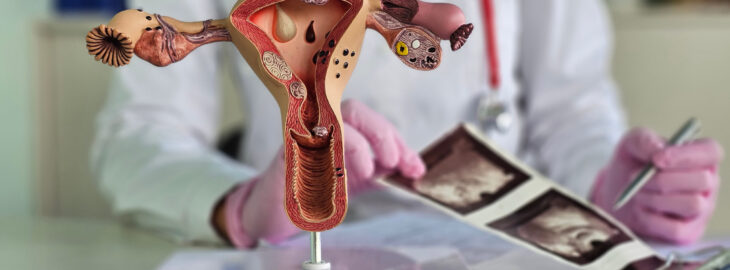
Recurrent pregnancy loss (RPL) is defined as the loss of two or more consecutive pregnancies of a viable fetus (2). Although different factors are associated with RPL, uterine abnormalities are thought to relate to around one in ten diagnoses.
The most influential guidelines published by ESHRE/ESGE, ASRM and Congenital Uternie Malformation by Experts (CUME) recommend the adoption of 3D ultrasound to evaluate the uterine factor in cases of RPL. However, the study by Busnelli et al is the first to report the prevalence of congenital and acquired uterine anomalies in RPL patients using 3D ultrasonography.
The authors found that the prevalence of uterine anomalies varied according to the diagnostic criteria used. For instance, partial septate uterus ranged from 29% to 60%, T-shaped uterus 3-4% and borderline T-shaped uterus approximately 4%. Furthermore, adenomyosis was detected in more than one in five (23%) patients and was associated with more severe forms of RPL, while FIGO type 0, 1 or 2 fibroids were detected in 4% of the included patients and endometrial polyps in 4% (1).
This variation in adopted diagnostic criteria and the role of adenomyosis in the RPL population, served as pivotal points for the ESHRE Journal Club discussion.
Since the study was a single-arm cohort, an important topic of discussion concerned the role of a control group in establishing the causal contribution of uterine factors in women with RPL. The Journal Club discussed that it is particularly challenging in retrospective studies to identify an appropriate control group of women with proven fertility, no history of abortions, and available 3D transvaginal ultrasound (TVUS) data.
Ideally, controls could be recruited from maternity clinics and include multiparous women (≥2 deliveries) who conceived within a defined time frame (6–12 months) and had no previous pregnancy losses. Conversely, it was suggested that women with at least two euploid pregnancy losses should comprise the RPL group. Any obvious etiology for RPL should serve as an exclusion criterion for both groups, while additional criteria can be adjusted according to the specific study design.
Although a recent multicentre study defined the sonographic criteria of “normal uterus” in nulliparous women, the Journal Club discussion concluded that uterine functionality cannot be fully confirmed before pregnancy (3). Ultimately, it was highlighted that the quality and cleanliness of the generated data are paramount, as compromising study design leads to random and difficult-to-interpret findings.
As pointed out by Busnelli et al, the prevalence of uterine anomalies, such as septate uterus, varies considerably depending on the classification system used, leading to significant discrepancies in diagnosis and management. For instance, an earlier study reported that while nearly a third (33%) of women were classified as having a septate uterus by at least one system, only 2.7% met criteria across all three, underscoring the inconsistencies between definitions (4).
Journal Club participants acknowledged that these variations increase the risk of both over- and under-diagnosis, potentially resulting in inappropriate treatment and complicating the interpretation of outcomes related to infertility and miscarriage. Thus, it was agreed that a unified classification system, developed through expert consensus, could help standardise practice and improve clinical relevance.
However, defining what constitutes a “normal” uterus remains challenging. Uterine morphology can change after pregnancy; and uterine functionality before pregnancy, such as implantation potential, is not yet fully understood. Moreover, it was pointed out that variability related to operator experience, imaging technology; and population differences further affects diagnostic consistency. Overall, it was concluded that these challenges emphasise the need for standardised, outcome-oriented criteria that focus on clinically meaningful measures rather than purely morphological definitions.
Journal Club experts and participants highlighted that diagnosing adenomyosis remains challenging due to its clinical and imaging overlap with conditions such as endometriosis, uterine fibroids, and heavy menstrual bleeding. This overlap often leads to misdiagnosis or delayed recognition.
The absence of a gold-standard diagnostic test further complicates detection. MRI and TVUS can miss early disease, and even 3D TVUS has limited specificity, despite a sensitivity of approximately 89%, leading to false positives. It was mentioned in the discussion that histological confirmation requires hysterectomy, which is not feasible in most reproductive-age women. Unfortunately, adenomyosis symptoms are often normalised by patients and clinicians alike and inconsistent use of standardised criteria, such as the MUSA classification, and low provider awareness can exacerbate variability in diagnosis.
Clinical outcomes appear to differ by adenomyosis subtype – focal, diffuse, interna, external – although data supporting this claim remains limited. Overall, Journal Club experts agreed that women with adenomyosis show higher miscarriage rates (35.4% vs 18.1% without adenomyosis) (5) and, as shown by Busnelli et al, a stronger association with recurrent pregnancy loss, particularly with three or more miscarriages (1). Given this variability, it was concluded that defining specific risk criteria is essential. Combining JZ measurements, multiple MUSA features, and a history of recurrent pregnancy loss could help identify high-risk patients and guide early intervention and monitoring. The cumulative live birth rate reaches approximately 60–70% in the ideal IVF setting where women younger than 35 years undergo multiple embryo transfers. This naturally raises the question: why not 100%? Could uterine factors represent the missing piece? Journal Club participants discussed that the reported prevalence of uterine factor infertility ranges from 2% to 16.7%, though estimates remain highly heterogeneous. Even with multiple embryo transfers, cumulative live birth rates vary between 68% after six transfers and 78% after ten transfers.
A Journal Club expert said that a progressive decline in success rates with each subsequent transfer would be expected if a fixed uterine defect were responsible for implantation failures. However, such a pattern is not observed. Consequently, it was highlighted that the reproducibility of uterine receptivity remains uncertain: a key determinant of residual IVF failure may be whether the uterus is consistently receptive or varies across cycles. As an in vivo assessment of uterine functionality is not yet available, it was concluded that existing evidence regarding the uterine factor should be interpreted with caution. To summarise, the uterine factor plays an important role in sustaining a pregnancy. Major guidelines on RPL recommend evaluating the uterus for congenital or acquired pathologies. Yet many questions remain regarding which specific structural characteristics of the uterus are associated with adverse reproductive and pregnancy outcomes.
Acknowledgements
The author would like to express his gratitude to all the members of ESHRE Journal Club who participated in this discussion (Noemi Salmeri, Attilio Anastasi, Juan J Fraire-Zamora and George Liperis) and the invited experts (Ursula Catena and Alexander M Quaas).
References
1 Busnelli A, Barbaro G, Pozzati F, D’Ippolito S, Cristodoro M, Nobili E, et al. The importance of the ‘uterine factor’ in recurrent pregnancy loss: a retrospective cohort study on women screened through 3D transvaginal ultrasound. Hum Reprod. 2024;39(8):1645-55.
2 Evaluation and treatment of recurrent pregnancy loss: a committee opinion. Fertil Steril. 2012;98(5):1103-11.
3 Gergolet M, Nicolì P, Vrtačnik Bokal E, Verdenik I, di Spiezio Sardo A, Zizolfi B, et al. Defining the “normal uterus” by ultrasound measurement of uterine lengths, thicknesses, and angles in a population of nulliparous women: the Normal UteRus asSEssment study. Fertil Steril. 2025.
4 Ludwin A, Ludwin I, Coelho Neto MA, Nastri CO, Bhagavath B, Lindheim SR, Martins WP. Septate uterus according to ESHRE/ESGE, ASRM and CUME definitions: association with infertility and miscarriage, cost and warnings for women and healthcare systems. Ultrasound Obstet Gynecol. 2019;54(6):800-14.
5 Cozzolino M, Cosentino M, Loiudice L, Martire FG, Galliano D, Pellicer A, Exacoustos C. Impact of adenomyosis on in vitro fertilization outcomes in women undergoing donor oocyte transfers: a prospective observational study. Fertil Steril. 2024;121(3):480-8.
You have to be logged in and an ESHRE member in order to comment.